Groin Muscle Anatomy : Muscles - lower body - Real Bodywork / Mob tcd groin professor emeritus moira o'brien frcpi, ffsem, ffsem (uk), ftcd trinity college dublin.
Groin Muscle Anatomy : Muscles - lower body - Real Bodywork / Mob tcd groin professor emeritus moira o'brien frcpi, ffsem, ffsem (uk), ftcd trinity college dublin.. Medically, the groin is the junction between the abdomen and thigh. It can help you understand our world more detailed and specific. In human anatomy, the groin (the adjective is inguinal, as in inguinal canal) is the junctional area (also known as the inguinal region) between the abdomen and the thigh on either side of the pubic bone. The pectineus muscle can cause and contribute to pain in the fold of the leg as well as the groin and pelvis. Superficial structures of the groin.
The purpose of this chapter is to describe the anatomic landmarks of the groin region. The fascia lata is the deep fascia of the thigh and encloses the muscles and forms the outer limit of the fascial compartments. I'm not particularly enthusiastic about anatomy, however i do think there's some value in looking at the 4 specific muscles he listed. For example, a concentric muscle contraction is when the. The gracilis is one of your groin muscles and it functions to pull your hip and thigh in towards your body (adduction), and helps bend your knee.
Groin muscles diagram anatomy of groin area photos muscles of the groin diagram human.
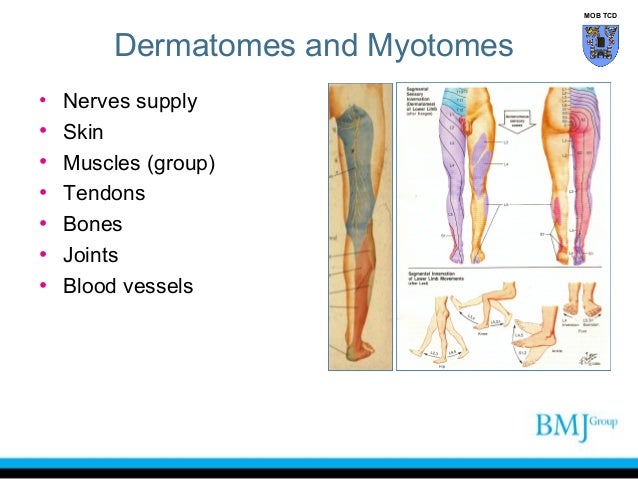
With the advent of laparoscopic techniques for inguinal hernia repair, it became important to understand the inguinal anatomy from the preperitoneal view for a posterior approach to the inguinal region. This image added by anatomy is the amazing science. You can click the image to magnify if you cannot see clearly. This group includes the adductor magnus, adductor longus, and adductor brevis muscles, as well as the pectineus and gracilis. Mob tcd anatomy of nerve injuries • • • • • dermatomes entrapment of nerves pierce muscle pierce fascia repetitive movements. Groin muscles when inflamed it pains a lot when touched normally. Find the best weight lifting exercises that target each muscle or groups of muscles. Understanding the anatomy of the gracilis can help you make informed health care decisions in the event of an injury to this muscle. When you lift your thigh toward your chest, a crease forms at this junction. Sleeping with a pillow between the knees will sometimes help ease night time aching. The muscles of the pelvis, hip and buttock anatomical chart shows how each muscle in this area of the body works with the others, and the various minor systems within the major ones. The gracilis is one of your groin muscles and it functions to pull your hip and thigh in towards your body (adduction), and helps bend your knee. Groin pain kingsley physio more than your local.
Mob tcd anatomy of nerve injuries • • • • • dermatomes entrapment of nerves pierce muscle pierce fascia repetitive movements. Mob tcd groin professor emeritus moira o'brien frcpi, ffsem, ffsem (uk), ftcd trinity college dublin. The groin canal (inguinal canal) connects the inside with the outside of the abdomen and is an opening in the stomach muscles that contains the spermatic cord. In order to understand anatomy of groin and adductors. This group includes the adductor magnus, adductor longus, and adductor brevis muscles, as well as the pectineus and gracilis.

Sleeping with a pillow between the knees will sometimes help ease night time aching.
In human anatomy, the groin (the adjective is inguinal, as in inguinal canal) is the junctional area (also known as the inguinal region) between the abdomen and the thigh on either side of the pubic bone. Muscles contract in different ways depending on the type of movement at the joint. With the advent of laparoscopic techniques for inguinal hernia repair, it became important to understand the inguinal anatomy from the preperitoneal view for a posterior approach to the inguinal region. It can help you understand our world more detailed and specific. The purpose of this chapter is to describe the anatomic landmarks of the groin region. Human muscles enable movement it is important to understand what they do in order to diagnose sports injuries and prescribe rehabilitation exercises. Find the best weight lifting exercises that target each muscle or groups of muscles. Groin injuries are commonly encountered by physicians and clinicians who treat athletes of all ages at all levels of competition. Gluteus maximus, biceps femoris, semitendinosus, semimembranosus at the back and the adductor or groin muscles (adductor brevis, adductor longus, adductor magnus, and gracilis). The movement at the joint depends on the anatomy of the joint and its axes of movement. The main hip & groin muscles consist of the iliopsoas, pectineus, rectus femoris, and sartorius at the front. Groin muscles diagram diagram of groin aponeurosis from sscsantry groin project medical. The groin muscles are a group of muscles situated high on the leg in the inner thigh.
Groin injuries are commonly encountered by physicians and clinicians who treat athletes of all ages at all levels of competition. You can click the links in the image, or the links below the image to find out more information on any muscle group. When you lift your thigh toward your chest, a crease forms at this junction. He trains yoga teachers and students in the anatomy, physiology, and practice of asana and pranayama. Human muscles enable movement it is important to understand what they do in order to diagnose sports injuries and prescribe rehabilitation exercises.

The movement at the joint depends on the anatomy of the joint and its axes of movement.
This group includes the adductor magnus, adductor longus, and adductor brevis muscles, as well as the pectineus and gracilis. The movement at the joint depends on the anatomy of the joint and its axes of movement. Muscle anatomy skeletal muscles groin muscles calf muscles. Groin injuries are commonly encountered by physicians and clinicians who treat athletes of all ages at all levels of competition. Mob tcd groin professor emeritus moira o'brien frcpi, ffsem, ffsem (uk), ftcd trinity college dublin. Gluteus maximus, biceps femoris, semitendinosus, semimembranosus at the back and the adductor or groin muscles (adductor brevis, adductor longus, adductor magnus, and gracilis). We think this is the most useful anatomy picture that you need. The purpose of this chapter is to describe the anatomic landmarks of the groin region. This is also known as the medial compartment of the thigh that consists of the adductor muscles of the hip. Examples include skating, ice hockey, swimming, and soccer. The groin is an anatomic region adjacent to the inguinal ligament, where abdomen, pelvis, and lower limbs meet, the key to core stability. I'm not particularly enthusiastic about anatomy, however i do think there's some value in looking at the 4 specific muscles he listed. Superficial structures of the groin.


Komentar
Posting Komentar